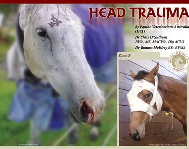
HEAD TRAUMA
by Dr Chris O'Sullivan and Dr Tamara McElroy (EVA)
 |
The Equine Veterinarians Australia (EVA) will be conducting clinics and demonstrations at Equitana Asia Pacific at the Melbourne Showgrounds Nov 20-23rd 2008. |
A blow to the head may appear to be nothing more than a surface injury yet, underlying damage, if severe, could have long-term effects on the horse's health and performance.
Head injury in horses can occur during accident or misadventure but it is typically a result of the head hitting either another object or the ground due to shying, bolting, pulling back, rearing up and over or a kick to the head from another horse. These types of injuries are seen more commonly in younger horses and are often associated with the breaking-in period, or as a consequence of paddock accidents. Head trauma requires rapid expert attention as, depending on the severity of the injury, it can result in damage to various structures including skin, bone, nerves, the eyes and the brain. The severity of damage, and the structures involved, will determine what treatment is required and the ultimate outcome for the horse.
Lacerations and grazes to the skin are often the most obvious signs of injury, however injuries to deeper structures may be less obvious initially. A horse with a head injury that has caused damage to the brain may show signs of changed behaviour, disorientation or become non-responsive to people or objects around them. In extreme circumstances the horse may be knocked out or suffer a seizure on the ground. The most important thing is to maintain a calm relaxed environment, call the vet and minimise danger firstly to the people around the horse and secondly to the horse. For example if the horse is seizuring on the ground the handler should not put themselves in a position to be injured by the horse, which is unaware that they are there, and padding should be placed around it to minimise the damage the horse may do to itself.
The veterinarian’s first step in assessing head trauma would be a thorough physical examination of the horse with a detailed inspection of the symmetry of the skull. This could include an oral exam to look for injury inside the mouth or failure of the teeth to normally line up (malocclusion) which may indicate a fracture of the jaw.
If trauma involves the eyes or surrounding area, a thorough examination of eyelid and eye function (an ophthalmologic exam) will be carried out to ensure the eye or surrounding structures are not damaged.
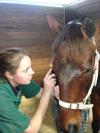
A neurologic assessment would also be performed, where the vet will look for signs such as head tilt, abnormal eye or ear position, blindness, muscle paralysis, depression or an altered gait. Identifying these abnormalities will help the vet to localise the most likely site of nerve or brain injury.
Once the initial assessment has been completed there are a variety of diagnostic tools available to further evaluate abnormalities identified should the veterinarian consider this necessary. Radiographs (X-rays) are valuable for locating and determining the presence and extent of bone fractures and whether the sinuses or brain cavity may be affected. Endoscopic examination (‘scope’) involves placing a long flexible tube up the horse’s nostril with a camera on the end to examine the nasal passages, including sinus openings and guttural pouches. This can provide important information regarding damage involving these areas and is particularly useful if blood is coming from one or both nostrils (epistaxis). The muscles that attach to the base of the brain cavity can also be viewed from the guttural pouches. Techniques such as a CT (Computed Tomography) scan or MRI (Magnetic Resonance Imaging) are becoming available, and in some cases can provide excellent detailed assessment of head injuries. However, these two techniques require general anaesthesia and the nature of many head injuries makes these horses unsuitable candidates.
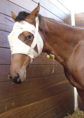
SKIN WOUNDS
Like most animals, horses have excellent blood supply to the head region and even extensive skin wounds heal well on the head compared with other sites on the body. Full thickness skin injuries to such areas as the lips and eyelids, however, if left untreated often heal with significant scarring, and may result in permanent deformity and dysfunction. Eyelid and lip lacerations, therefore, heal best when repaired surgically.
FRACTURES
Hits to the head which don’t cause bone fractures - a break in the bone - can result in a bony lump, commonly known as a callus, forming 4-6 weeks after the injury between the bones of the skull. This is seen most commonly across the forehead between the eyes.
Skull fractures can be divided into fractures of the jaw bone (mandible), brain cavity (cranium), and the other bones of the head including forehead, sinuses and orbit bones surrounding the eye. Jaw fractures are most easily identified by a horse having difficulty eating, dropping food, drooling or misalignment (malocclusion) of the teeth. Treatment options depend on the location of the fracture within the jaw, but in most cases jaw fractures should be repaired surgically. A variety of techniques may be applied to repair the fracture including metal plates, wires, intraoral splints (like human braces and plates), or external fixation techniques (pins screwed into the bone through the skin and attached to a rod).
Facial bone fractures vary in severity, from a mild facial deformity to more severe open fractures entering the sinuses, distorting the nasal cavities, or impinging on the eye ball. With severe fractures of the nasal cavity, horses can develop trouble breathing, particularly as swelling occurs after the accident. These horses may require a tracheostomy (a temporary hole in the wind pipe) to enable them to breathe properly. Though this may seem alarming to a worried owner, it’s a simple and effective procedure.
If a cosmetic result is not important for the injured horse then those fractures that have not broken the skin (closed fractures) can often be left untreated. Surgery is advised for more severe or ‘open’ wound fractures, where an improved cosmetic result is desired, and in fractures that may affect the eye or airway functions. Any fractures with exposed bone or involving the sinuses carry a high risk of infection.
Fractures of the cranium (top of the head and surrounding the brain), typically occur as a result of the horse flipping over backwards or rearing and striking the poll, usually resulting in direct damage to the brain. These fractures most commonly occur in the skull at the base of the brain, and are not obvious when looking externally at the head, therefore good quality radiographs are required to identify them. Horses with these injuries may have bleeding from the nose and or ears, and unfortunately, in injuries of this nature there will usually be mild to severe brain or neurological damage.
INJURY TO THE NERVOUS SYSTEM
The most obvious sign of serious neurological or nervous system injury is the horse experiencing a loss of consciousness or uncontrolled seizures. Horses in a coma, or seizuring, who fail to respond to veterinary treatment within 24 hours of injury, are highly unlikely to recover. This category of horses require rapid intensive care treatment and may be treated with many different drugs including anti-inflammatories, anti-seizure drugs, antioxidants and diuretics. If the horse does respond within 6-8 hours there is a favourable prognosis for life, but the ultimate usefulness of the horse may not be determined for many months, as those that recover initially may go on to deteriorate several months later if fracture-healing-callus impinges on nervous tissue.
When the middle ear system (vestibular) is damaged, the horse’s balance and coordination can become affected and it may walk in circles, lose it’s balance or develop a head tilt. Though this is distressing for the owner to see, the animal will often respond well to treatment, and it learns to visually compensate as healing progresses. Affected horses often compensate better if kept outside, rather than stabled since it allows them to be more active and learn to adjust. Horses with skull base fractures or vestibular syndrome who fail to improve within the first couple of days have a poor prognosis as performance athletes.
The nerve that controls the muscles of expression in the head and face exits the skull base and runs along the side of the head. Damage to this nerve will result in drooping lips and nostrils, facial palsy and the inability to blink or close the eye. This inability can rapidly result in drying of the cornea, ulceration and ultimately loss of the eye. The good news is that the majority of facial nerve injuries will gradually regain function, but it can take a prolonged period of time (3-6 months or longer). Unfortunately, there will always be some of these horses that will not regain these functions.
Head injuries are as potentially dangerous and life-changing for horses as they are for humans and, because of the nature of horses, will continue to occur despite good horse keeping practices or safe accommodation. When head trauma does occur or is suspected, the accurate identification of specific structures involved and the severity of damage is of paramount importance. Rapid and thorough clinical, ophthalmologic (eye), oral and neurologic examinations, combined with appropriate advanced diagnostics, give the best opportunity for correct treatment and a more positive outcome for the head injured horse.
|
Dr Chris O’Sullivan (BVSc, MS, MACVSc, Dip ACVS)
A registered specialist equine surgeon at Randwick Equine Centre, Chris
graduated from Sydney University and trained in surgery at The Ohio State University USA. He enjoys working with performance horses and his specific areas of interest include lameness, diagnostic imaging (Computed Radiography, Ultrasonography, Scintigraphy), orthopaedic and soft tissue surgery.
Dr Tamara McElroy (BSc BVMS)
A first year graduate from Murdoch University in Western Australia, Tamara is currently an intern at Randwick Equine Centre.
Randwick Equine Centre is a seventeen veterinarian purely equine practice servicing the greater Sydney area. The hospital facilities include a modern surgery, laser surgery, radiology (x-ray), digital radiology, ultrasound, shock wave therapy, video-endoscopy (airway and stomach) and nuclear scintigraphy. See the web site: www.randwickequine.com.au |
| |
|
|