|
Vol 36-6 April May 2015
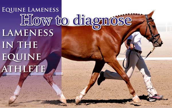
Diagnosing Lameness in the Equine Athlete
by Dr Max Hall
 BVSc(Hons) MANZCVS(Equine Surgery) Diplomate ECVS(Equine Surgery) BVSc(Hons) MANZCVS(Equine Surgery) Diplomate ECVS(Equine Surgery)
Maxwell is a Diplomate of the European College of Veterinary Surgeons and is a
specialist in equine surgery. He currently resides in Perth, working between WA and
one of N.Z’s busiest equine practices - Cambridge Equine Hospital - and Goulburn
Valley Equine Hospital, Victoria. Maxwell has a particular surgical interest in
orthopaedics and upper respiratory tract surgery.
Being able to tell if a horse is lame and determining where the problem originates can be difficult at times for even the experienced horse person. Not all lamenesses appear as a horse ‘limping’, some may just be a bobbing of the head, a change in the way the horse carries itself overall, or just a change in attitude. Many riders can ‘feel’ something is just not right with their horse, often before there is visually a noticeable difference in movement, and early intervention by a veterinarian can often prevent further damage.
To be able to identify and understand lameness in horses, which can involve virtually any part of the body and can originate in bone or soft tissue, riders and owners must know how to distinguish between normal and altered movement.
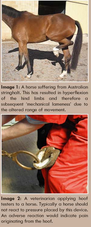
Lameness is defined simply as an abnormality in a horse’s movement caused by pain or reduced range of motion (see image 1) and the term covers a wide range of ailments. A horse that is lame is often referred to as being unsound.
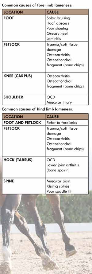
The most common health problem seen in the equine athlete; as a general rule 75% of lameness issues are seen in the forelimb whilst the remainder are hind limb issues. Of the lameness problems seen in the forelimb 95% of these originate from below the knee or hock. Some horse owners think of lameness only as a problem of the feet or legs and never look further than these areas, however, the most commonly misdiagnosed lameness issues in horse are suspected shoulder and hip lamenesses. It is always a wise idea to start by examining the hoof initially and then looking for other explanations.
Locating the source of lameness and then developing a plan to manage it are high priorities and these procedures will generally involve the veterinarian or farrier, or often both.
How to determine if a horse is lame:
A lame horse is in pain. As an owner it is your responsibility to be able to tell if the horse is lame and then seek appropriate treatment. When veterinarians are assessing lameness, horses are commonly graded from 0-5 based on the level of the lameness.
This scale indicates the severity of the lameness. This 0-5 scale had originated from the AAEP (American association of Equine Practitioners) system and is as follows:
Lameness not perceptible under any circumstances.
Lameness is difficult to observe and is not consistently apparent, regardless of circumstances.
Lameness is difficult to observe at a walk or when trotting in a straight line, but consistently apparent under certain circumstances (e.g., weight carrying, circling, inclines, hard surfaces, etc.)
Lameness is consistently observable at a trot under all circumstances
Lameness is obvious at a walk
Lameness produces minimal weight bearing in motion and/or at rest or a complete inability to move.
Terms such as ‘acute’ and ‘chronic’ are sometimes used when assessing lameness. Both terms are used to explain the duration of the condition. Acute means that the condition has occurred suddenly and has not been present for a long period of time. Chronic conditions are of long-standing duration that may have been present for several weeks to months. The severity of the lameness (see above) is an independent scale to that of the duration.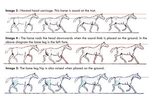
Other important variables of lameness are whether it is persistent or intermittent, and progressive or static. There is uncertainty amongst horse owners with these terms, some confusing persistent with chronic, but chronic refers to the length of time of the problem while persistent means that the lameness (which could be recent or not) has been consistently observable since its onset – not coming and going (e.g. intermittent).
Unless the horse is non-weight bearing (5/5 lame) or lame at the walk (4/5 lame) lameness is most easily assed while the horse is trotting. To fully recognise the extent of lameness the horse should be observed at the trot in a straight line and on circles both ways while on the lunge. This should be done with the horse being worked on both a hard and soft surface that is even and free of debris, as an area strewn with debris is not going to give a true indication of the horse’s lameness. 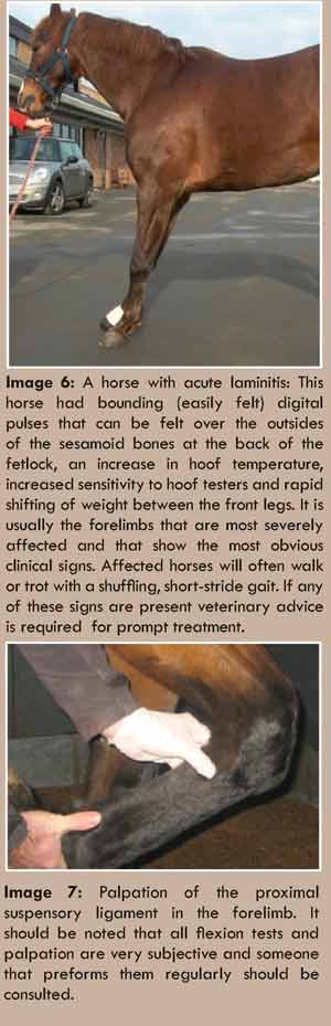
Many high-speed lameness problems are only present when the horse is cantered therefore the horse should also be viewed in this gait if appropriate. If the pain is only in one leg it should be easy to notice that the horse is not moving evenly. Some horses can be lame in multiple limbs – which makes diagnosis much more difficult.
A sound horse should move evenly in its stride with minimal head movement. The hips should rock evenly behind with no toe drag along the ground. (see image 3)
A lame horse will often throw its head in rhythm with its stride. If the horse is sore in a front leg, it will throw its head up as the sore side touches the ground or down when the sound leg hits the ground (see image 4 ). ‘Head nodding’ is typically associated with forelimb lameness.
If the lameness is in a back leg, the horse will lean (the hip) onto the sound side (down on sound). He may also drag the toe of the lame limb. (see image 5). Hip drop. If the horse is painful in both front feet (or hind) it will typically move with a very shuffled gait and only be obviously lame when lunged in either direction.
 A quick look as the horse wanders in for its morning feed or when it is turned out later in the day can tell horse owners a great deal about the horse’s health and physical condition. A reluctance to move or an unwillingness to trot out should raise warning flags that all is not well. The stance of a horse when resting is also important to assess. It is common for a normal horse to relax a hind limb off the ground and then switch onto the other limb. A horse pointing out a front limb, or even leaning back off its feet can be suggestive of pain (see image 6) A quick look as the horse wanders in for its morning feed or when it is turned out later in the day can tell horse owners a great deal about the horse’s health and physical condition. A reluctance to move or an unwillingness to trot out should raise warning flags that all is not well. The stance of a horse when resting is also important to assess. It is common for a normal horse to relax a hind limb off the ground and then switch onto the other limb. A horse pointing out a front limb, or even leaning back off its feet can be suggestive of pain (see image 6)
Once it has been determined that the horse is actually lame, what is the next step?
Always check the foot! Hoof abscesses are also known as ‘the great pretender’ and due to the level of pain (severe) being displayed by the animal they are commonly mistaken as a fracture. While more common in the front feet they do occur in hind hooves.
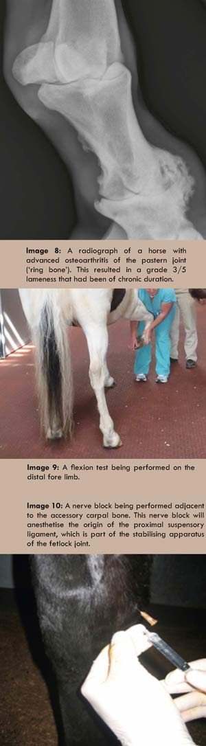
Depending on the level of lameness the veterinarian and or farrier will conduct an initial examination that would include an accurate medical history, full physical examination and a more specific lameness examination. If your veterinarian has been called out it is likely he or she will palpate (see image 7 ) the affected limb and even perform flexion tests to try and isolate the lameness. Things they will be feeling for are increased heat, swelling and pain of specific areas. Flexion tests are when an area of the horse’s limb is held in ’flexion‘ for a set amount of time and then the horse is trotted off. A positive response is when the horse becomes significantly lamer after the test.. (see image 9 )
In these illusive cases nerve blocks can be performed. Nerve blocks (see image 10) are when the nerve or nerves to a specific anatomical location are desensitised by infusing local anesthetic into the area. They can be performed on most areas of the front and hind limb and can be extremely helpful in identifying the area of pain. A horse will typically become sound once local anesthetic has been applied to the location causing pain (e.g. the fetlock joint). The appropriate diagnostic modality (e.g. ultrasound or radiography) can then be used to focus on and further assess the area.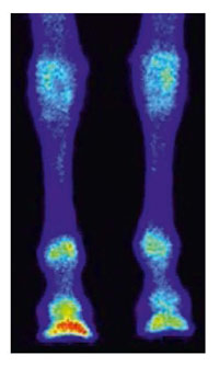
Occasionally lamenesses are not a leg issues and the pain is originating from anywhere from the back (e.g kissing spines – see image 11), head, neck, or pelvis. These cases can be very complex and apart from nerve blocks some other more advanced diagnostics tests are required. Nuclear scintigraphy (aka ‘bone scanning’) can be used to assess structures such as the neck and pelvis more easily.
Lamenesses can become extremely complex and occasionally involve multiple limbs, however this article is aimed at giving a basic understanding on how lameness can be detected and then localised to a specific area. Involving a veterinarian early once a lameness is noticed can significant improve the chances of the horse becoming sound, as appropriate treatment can be undertaken promptly.
 |